Bone Health and Hormones
My bone health wake-up call
In my late twenties, I discovered I had reduced bone mineral density (osteopenia) in my spine. This followed two years of amenorrhoea (missing periods), multiple years on the combined oral contraceptive pill and a history of chronic stress, undernutrition, and over-exercise. There is perhaps a beautiful irony in how this discovery proved a turning point in my own recovery. The same is true for many women I am now privileged to support. Whilst treatments to reverse functional hypothalamic amenorrhoea (FHA, often referred to as HA) are vital for bone health, the deleterious effects on bone often provide the wake-up call needed to ignite change and put women back in control of their health.
Contrary to popular belief, bone is more than just scaffolding for the body. The health and metabolism of bone are complex and governed by many factors, including hormones, genetics, nutrition, lifestyle and more.
This blog post and the next aim is to provide an insight into the various factors that influence the health of our bones. This one looks mostly at hormonal influences (of which there are many!) and part two will take a deeper dive into the nutrition and lifestyle aspects of bone health.
What is Bone?
Bones are often thought of as static, inert structures which only offer structural support. However, like other organs, bones have many functions. Bones enable movement, protect vital organs, and even act as a storage site for certain nutrients including calcium, phosphate, and magnesium. Recent studies have shown that bone cells also produce hormones (FGF23 and osteocalcin) that are involved in mineral and energy balance [1].
Bone is a dynamic tissue that is continually remodelled by the action of two different types of bone cells, namely osteoclasts and osteoblasts. Osteoclast cells mediate the resorption of bone (a process which involves the breakdown of bone tissue) and work alongside osteoblast cells which encourage bone formation. Bone resorption and formation are normally tightly controlled within the body to maintain a net bone mass; a dynamic balancing act often referred to as bone homeostasis. Any disturbance to bone homeostasis (causing an imbalance in bone resorption and formation) can give way to health consequences including osteoporosis [2].
Peak Bone Mass and Bone Density
Bone mass increases progressively throughout childhood, with peak bone mass (PBM) defined as the maximum bone mineral density (BMD) obtained during development. Humans gain the majority of their PMB during adolescence; however, bone continues to increase in density in early adulthood peaking at around 25-35 years in women [3].
Achieving peak bone mass has wide-ranging benefits to both physical and mental health. An increase in PBM by one standard deviation has been shown to reduce fracture risk by 50% in later life [4]. What’s more, failure to achieve PBM increases the risk of osteoporosis, a condition characterised by low bone density, reduced bone strength and increased fracture occurrence [5].
Low bone density has also been shown to impact psychological wellbeing, with osteoporotic patients commonly experiencing feelings of anxiety, fear of falling and depression. Loss of independence and reduced self-esteem are also reported amongst those with osteoporosis [6].
Risk Factors for Low Bone Density
Several factors can disrupt the normal physiology, integrity, and health of bone, including nutritional status, body weight, endocrine (hormone) disorders, genetics, medications and/or chronic disease [3]. Since adolescence and early adulthood are crucial times for bone development, any disturbance to bone homeostasis during these periods is particularly significant and can jeopardise the achievement of full genetic potential PBM.
Hormones
Numerous hormones are involved in the growth of the skeleton and bone mass accrual including sex hormones (oestrogen, progesterone, and testosterone), calcium regulating hormones (parathyroid hormone, calcitriol, and calcitonin), growth hormone, thyroid hormones, stress hormones and others. These are discussed in more detail below.
Body weight
Low BMI has been identified as a major risk factor for low bone mineral density and is a predicting factor for accelerated bone loss in older age. This is partly owing to the physical and mechanical stress that body weight places on bone. A heavier physique is more demanding on the skeleton which naturally strengthens and adapts to carry the load.
Energy restriction and weight reduction also impact levels of several hormones involved in bone regulation, including sex hormones and leptin. Additionally, reduced fat mass can independently decrease circulating oestrogen and other sex hormones, negatively impacting osteoblast and osteoclast activity [7].
Physical activity
A large body of evidence supports claims that weight-bearing exercise has a positive impact on bone mineral density in healthy populations [8]. However, when combined with low caloric intake and undernutrition, exercise can become a risk factor for hormonal disturbances, including functional hypothalamic amenorrhoea and hypogonadism, both of which can result in bone loss [9]. You can read more about this concept in this blog post.
Stress
Emerging evidence suggests that poor mental health may precede bone loss. In a 2017 study, mental health problems (including psychological stress, depression, and suicidal ideation) were associated with lower BMD in both pre-menopausal women and men [10].
Psychological stress activates the sympathetic nervous system (fight or flight response), hypothalamic-pituitary-adrenal (HPA) axis and promotes the secretion of stress hormones, including cortisol. Excess cortisol has been suggested to trigger FHA by inhibiting the frequency of gonadotropin releasing hormone (GnRH) pulses and inhibiting reproductive function at the hypothalamus, pituitary, and uterine levels [11]. One of the consequences of this is reduced gonadal hormones (oestrogen, progesterone, and testosterone) with subsequent implications for bone health.
There is evidence to suggest that psychological stress can also directly impair bone health. Circulating stress hormones can suppress growth hormone, inhibiting osteoblastic bone formation, and trigger the release of inflammatory cytokines. Increased inflammation associated with stress has been associated with increased bone resorption via upregulation of surface receptors on osteoclast cells [12]. Together, these effects are linked to net bone loss [13].
Nutrition
Adequate energy availability is essential for bone homeostasis. Short-term low energy availability has been shown to increase bone resorption and decrease bone formation amongst physically active women, whilst longer-term low energy availability is associated with lower bone mass, reduced bone strength and increased risk of fracture and osteoporosis [14].
In addition to energy intake, numerous nutrients play a role in bone health. Adequate protein and fat intake are vital for promoting optimal bone health. Most of us recognise that adequate calcium intake is important for bone, but other micronutrients including vitamin D, phosphorus, magnesium, vitamin K2, and vitamin C are also essential. These will be discussed in more detail in the next blog post.
Malabsorption (gut) disorders and other medical conditions
Patients with gastrointestinal diseases (e.g., coeliac disease, pancreatic insufficiency, inflammatory bowel disease and chronic liver diseases), tend to be at increased risk of osteoporosis. Bone issues may also arise secondary to treatments for gastrointestinal conditions, including bowel surgery and/or the use of certain medications.
Malabsorption of nutrients because of these disorders increases the likelihood of deficiencies in important bone nutrients, including calcium, phosphorous, vitamin D, vitamin K and even protein, leading to increased rates of bone loss. Elevations in cytokines associated with inflammatory conditions may also directly impact bone homeostasis. Medications known as protein pump inhibitors (PPIs) that reduce gastric acidity may also decrease calcium absorption [15].
Medications
As well as PPIs, many other common medications have been suggested to have harmful effects on bone health including glucocorticoids, selective serotonin receptor inhibitors (SSRIs), thiazolidinediones, anticonvulsants, medroxyprogesterone acetate, aromatase inhibitors, androgen deprivation therapy, heparin, calcineurin inhibitors, and some chemotherapies [16]. If you take any of these medications and you are concerned about your bone density, you may want to talk to your health provider about alternatives or how you can increase certain nutrients to counteract side-effects that may affect bone health.
Smoking & Alcohol
Other lifestyle factors including smoking and alcohol consumption can have detrimental impacts on bone.
Tobacco smoking can indirectly affect bone health, through suppression of appetite and weight loss, disruption to the parathyroid-vitamin D axis, adrenal and sex hormones, as well as reducing oxygen supply to bones. Tobacco smoke also has direct impacts on bone tissue by inhibiting bone synthesis and angiogenesis (the formation of new blood vessels) in bone [17].
Similarly, chronic alcohol consumption is correlated with lower BMD. The mechanisms behind this observation remain unclear due to the frequent presence of confounding factors such as alcohol-related comorbidities, age and other lifestyle factors. However, the observed effect to BMD may be attributed to malnutrition as well as alterations in hormone levels and signalling [18].
The Influence of Hormones – a closer look
As mentioned above, there are several hormones involved in bone homeostasis. These include sex hormones (oestrogen, testosterone, and progesterone), calcium-regulating hormones (parathyroid hormone, calcitriol, and calcitonin), and other hormones such as growth hormone, thyroid hormone, cortisol, insulin, and leptin [19]. What follows is a detailed overview of how some of these hormones are involved in regulating bone health.
Sex hormones
Oestrogen
Oestrogen is the main hormone regulator of bone metabolism in both men and women. The fact that osteoporosis is four times more prevalent in women [20] may be due in part to menopause, a time where normal bone homeostasis is impaired by oestrogen deficiency.
Despite the acknowledgement of the importance of oestrogen in bone regulation, the mechanisms remain unclear. However, research continues to develop an understanding of how oestrogen inhibits bone resorption and initiates bone formation.
Studies have consistently identified osteoclasts as a direct target for oestrogen, which plays a role in programming the death of these cells (apoptosis), blocking transcription of molecules regulating their activity, and suppressing their development [21]. Oestrogen has also been suggested to indirectly affect osteoclast formation and activity via other cells (osteoblasts, T cells and B cells) and regulate the production of some bone-resorbing cytokines.
Overall, the effect of oestrogen on osteocytes (established bone cells), osteoclasts, and osteoblasts is thought to lead to inhibition of bone remodelling, decreased bone resorption, and maintenance of bone formation, explaining the protective effects of oestrogen on the skeleton and why declining levels of oestrogen (during FHA and menopause) leads to dysregulation of bone homeostasis.
Testosterone
Testosterone is the major sex steroid in males but is also produced in small amounts by the ovaries in females. Through the action of an enzyme known as aromatase, testosterone can be converted to oestradiol (the strongest form of oestrogen) [22]. Thus, normal levels of testosterone may contribute to the bone protective effects of oestrogen mentioned above.
Additionally, circulating androgens (male sex hormones, including testosterone) have their own direct influence on bone through interaction with androgen receptors on bone-forming cells. The presence of testosterone appears to stimulate the proliferation of osteoblasts and inhibit their apoptosis (cell death) [23]. What’s more, androgen deficiency may lead to the proliferation of osteoclasts (bone-resorbing cells).
Progesterone
Progesterone is another major sex hormone in women and has also been found to have bone protective effects. The mechanisms behind these effects are not yet understood, but emerging evidence indicates progesterone may stimulate osteoblast differentiation, favouring bone formation over resorption [24].
Calcium-regulating hormones
Most of us are aware of the importance of calcium for bone health, with calcium salts constituting the majority of bone tissue’s inorganic mineral component. So, it may come as no surprise that calcium-regulating hormones play an important role in bone homeostasis.
Parathyroid hormone (PTH) and Calcitriol
Produced behind the thyroid gland, PTH tightly controls calcium levels in the blood. The parathyroid gland is sensitive to small changes in the blood concentration of calcium and so when calcium levels drop PTH secretion increases.
PTH works by increasing calcium absorption in the gut, reducing loss of calcium through the kidneys and increasing renal production of another hormone, calcitriol (the most active form of vitamin D). Similarly, the main action of calcitriol is to increase the intestinal absorption of calcium and phosphorus [19].
PTH and calcitriol also work together to stimulate osteoclastic bone resorption and the release of calcium from bone to blood. Withdrawals can be made from the bone “bank account” to enable the body to closely regulate blood levels of calcium. Whilst this is a physiologically normal and essential process if the parathyroid glands produce too much PTH (a medical condition known as hyperparathyroidism) excess calcium can be released from bone stores with consequences for bone integrity.
Calcitonin
In contrast, the main action of calcitonin, which is produced by the thyroid gland, is to lower calcium levels in the blood. One of the ways it does this is by inhibiting the action of osteoclasts and thus blocking the breakdown of bone [19]. For this reason, calcitonin is sometimes used as a drug for the treatment of bone diseases.
Other hormones
As well as the sex and calcium regulating hormones, other hormones are also involved in bone homeostasis. These include:
Growth hormone (GH)
Released from the anterior pituitary gland, GH is a crucial regulator of longitudinal skeletal growth in children.
GH exerts direct action on osteoblasts via GH receptors, stimulating osteoblast proliferation and differentiation.
GH also stimulates the production of insulin-like growth factor 1 (IGF-1).
IGF-1 promotes the production of osteoblasts and reduces their apoptosis (cell death).
Therapeutic administration of both GH and IGF-1 has a positive effect on osteoporosis and fracture healing [25].
Thyroid hormone
Deficiency has been associated with impaired growth in children.
Thyroid hormones are important for increasing energy production and regulating metabolic activity in all body cells, including bone cells.
Normal thyroid function is essential to bone homeostasis and normal rates of bone formation and turnover [26].
Cortisol
Released from the adrenal gland, cortisol is involved in the stress response and aids the body’s reaction to stress and injury.
The influence of cortisol on the skeleton is complex. Small amounts of cortisol are needed for growth and development of bone, but large amounts of cortisol have been found to inhibit bone growth.
Cortisol has been suggested to impair the synthesis of osteoclasts and osteoblasts and increase cell death (apoptosis) of both cell types.
Overall, cortisol leads to negative bone turnover and increases bone loss and fracture risk [27].
Insulin
Emerging evidence suggests that insulin may have anabolic effects on bone by stimulating the differentiation of osteoblast cells.
It is not yet clear whether the effect on bone is the direct action of insulin, or whether it is the observed effect of increased muscle work and skeletal loading [28].
Leptin
Emerging evidence suggests that leptin may promote bone formation.
Leptin receptors have been found on osteoblast cells, suggesting a direct impact on bone health.
Leptin is involved in the regulation of osteocalcin (which in turn regulates bone metabolism as well as insulin sensitivity and energy expenditure).
In human studies, it is difficult to distinguish between the effects of leptin and the effects of other hormones that increase with leptin therapy, including oestrogen, cortisol, IGF-1 and parathyroid hormone [29]. As such, research into the mechanisms behind leptin and bone health is still evolving.
Functional Hypothalamic Amenorrhoea and Bone Health – what’s the link?
Functional hypothalamic amenorrhoea (FHA, or HA for short) is a disorder often arising and associated with chronic stress, over-exercise, and undernutrition. HA is characterized by irregularity in GnRH pulsatility, suppression of the hypothalamic-pituitary-ovarian (HPO) axis and chronic anovulation. For this reason, oestrogen deficiency is a symptom of HA.
As described above, oestrogen plays a key role in bone homeostasis and thus low oestrogen (as in HA) can result in both the failure to achieve peak bone mass and bone loss, both of which increase fracture risk and the chances of developing osteoporosis in later life. However, it’s also important to appreciate the myriad potential causes and hormonal aberrations that characterise this complex condition. In fact, HA is associated with a wide range of other hormonal disturbances that may negatively influence bone integrity, including those discussed in this article. Consequently, treatments to reverse HA are vital for bone health.
Guidelines for the treatment of HA include restoring weight, reducing excessive exercise and stress management. Nutritional rehabilitation is also a cornerstone of recovery for most clients I work with. Additionally, current medical guidelines recommend considering hormone replacement therapy (HRT) should amenorrhoea persist for 12 months or more [30, 31].
Hormonal Contraception, Hormonal Replacement Therapy (HRT) and Bone Health
Recognition of the impact of hormonal deficiency on bone health has led to the development of many studies assessing the efficacy of oral contraceptives and other hormone replacement therapies on bone mass. Recent systematic reviews and meta-analyses of available evidence suggest that evidence is limited for the use of oral contraceptives and hormone replacement therapy in pre-menopausal women.
A 2006 systematic review [32] studied the use of the oral contraceptive (OC) pill on bone mineral density in 4 groups of women (healthy pre-menopausal, oligo/amenorrhoeic pre-menopausal, anorexic pre-menopausal and perimenopausal). The study concluded that there was ‘good’ evidence for the use of OC in perimenopausal women, ‘fair’ evidence for oligo/amenorrhoeic women and ‘limited’ evidence for both anorexic and the healthy pre-menopausal groups. The latter conclusion was explained by the idea that women with anorexia may be more sedentary than other groups and thus oestrogen deficiency played less of a role (and insulin-like growth factor 1 more of a role) in reduced bone mineral density. Interestingly, the study found that amongst the healthy pre-menopausal group, a combination of exercise and OC use may negatively impact bone mineral density, which is a significant finding for exercising women. This finding was proposed to be due to nutritional calcium deficiency, or due to suppression of internal hormone release.
Conversely, a 2017 systematic review and meta-analysis [33] found a significant increase in bone mineral density at the lumbar spine in patients with HA after receiving OC for 12 months. Bone mineral density at other sites in the body was unchanged and overall increase in BMD was not significant compared to placebo. The researchers also admitted that the quality of evidence was low due to a high risk of bias in this study, combined with its small sample size and indirectness of measures.
Interestingly, a 2007 systematic review of the literature surrounding the use of various hormonal replacement therapies (HRT) for bone loss in HA [34] found inconsistent improvements in bone mineral density. HRT (including oestrogen and androgen replacement) led to no improvements in bone mineral density in women with HA when compared to placebo. The researchers concluded that although bone resorption may be suppressed by the replacement of these hormones, there seems to be a greater suppression of osteoblast activity and a resulting inability to replace resorbed bone. On the other hand, increasing dietary energy intake and resultant weight gain and/or resumption of menses were found to increase bone mineral density by 1.1-16.9% and this was concordant with an increase in bone formation and decreased markers of bone resorption.
Finally, a systematic review published in 2021 examined the effects of various estrogen replacement therapies (oestrogen oral contraceptives, conjugated oestrogens, and transdermal estradiol) on BMD in premenopausal women with HA [35]. In agreement with the other studies, this review found no evidence to support the use of oral contraceptives or conjugated oestrogens for improving BMD. This study was the first to review the use of a transdermal oestrogen patch with this patient group, and data suggested that this treatment led to a significantly increased spinal BMD. The theory behind this is that, unlike OC, administration of oestrogen through the skin bypasses hepatic (liver) first-pass metabolism and so does not lead to downregulation of IGF-1 (which we know is a bone-promoting hormone).
Combined, these findings suggest that whilst hormonal therapies including HRT and OC may be effective in decreasing further bone loss, they appear ineffective at promoting bone formation. Nutritional therapies aimed at restoring energy balance show a stronger positive effect on bone health, and thus should be attempted as a first-line initiative. Transdermal oestrogen patches could be considered a second-line initiative to optimise bone health [31].
When to Take Action
So, how do you know when to act and what investigations can you consider? If you are concerned or curious about your bone health, my advice is to consider speaking to your GP or another qualified and registered health specialist, if:
Your period has been missing for 3 months or more. You don’t take hormonal contraception and you are not pregnant
Your period has stopped for 3 months or more whilst taking hormonal contraception, having previously had a regular withdrawal bleed
You have experienced one or more stress/bone fractures in the last 12 months
You have a low body weight or a history of low body weight
You engage in food restriction with or without exercise dependence/addiction
You have a malabsorption-related gut disorder (e.g., coeliac disease or inflammatory bowel disease)
You have a history of chronic stress or consider yourself anxious or depressed on a regular basis and have one or more additional risk factors for low bone density
You are concerned about any of the risk factors outlined in this blog and/or you have a family history of osteoporosis, bone injury or fracture
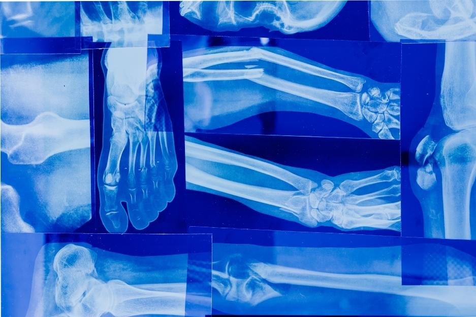
First-line testing for bone density is a DEXA scan. This scan uses low-dose X-rays to measure the amount of radiation that can pass through bone, helping to assess the risk of bone diseases including osteoporosis. The result is provided as a ‘T-score’ which represents how an individual’s bone density compares to average peak bone density (a score of -1 or above is considered ‘normal’). Private testing companies can also be used if the test is not available to you on the NHS.
Additional tests to consider include a comprehensive sex (pituitary and ovarian) hormone assessment, thyroid hormones, cortisol and other adrenal hormones (preferably assessed across the day as a dried urine or saliva test), bone profile and vitamin D.
It’s worth noting that a calcium blood test will not measure whether you are getting enough calcium in your diet or whether you might need a supplement. This is because your body works hard to keep calcium levels within a required range in the blood. Checking for low or high calcium levels, either using a blood or urine test, is generally more useful to indicate whether you have an underlying medical condition that is affecting your bones.
For more advice on any of these tests, including private DEXA scans, blood tests and more, you are welcome to contact me or set up a short free call so we can discuss your needs together.
Take-home messages
Bone is a dynamic tissue that is continually remodelled.
Bone remodelling must be tightly controlled to optimise peak bone mass and prevent bone loss.
Achieving optimal peak bone mass has numerous physical and mental health benefits.
Risk factors for low bone mass include hormonal disruptions, low body weight, low physical activity or unsupported excessive exercise, stress, sub-optimal nutrition, malabsorption, certain medications, smoking and alcohol use.
Several hormones are involved in the regulation of bone homeostasis including sex hormones, calcium-regulating hormones, growth hormone, thyroid hormone, cortisol, insulin, and leptin.
Individuals with HA are primarily at risk of low bone mass due to hormonal disruptions including oestrogen deficiency. Other contributing factors may include low body weight, low body fat %, malnutrition and stress.
There is limited evidence for the use of oral contraceptives in optimising bone health in HA. Optimising nutrition and restoring energy balance should be attempted as a first-line initiative, whilst transdermal oestrogen patches may be considered as a second-line initiative to optimise bone health.
If you are concerned or curious about your bone health, speak to your GP or another qualified and registered health specialist.
As always, it’s also important to remember that there is no one approach to optimise bone health that works for everyone. If you'd like to find out more about what you can do to support bone health, please book a free call so we can discuss your particular needs.
REFERENCES
1. Zhou R, Guo Q, Xiao Y, Guo Q, Huang Y, Li C, Luo X. Endocrine role of bone in the regulation of energy metabolism. Bone Res [Internet]. 2021 May [cited 2023 Aug 7]; 9 (1): 25. Available from: https://pubmed.ncbi.nlm.nih.gov/34016950/ doi: 10.1038/s41413-021-00142-4.
2. Florencio-Silva R, Sasso GR, Sasso-Cerri E, Simões MJ, Cerri PS. Biology of Bone Tissue: Structure, Function, and Factors That Influence Bone Cells. Biomed Res Int [Internet]. 2015 Jul [cited 2023 Aug 7]; 2015: 421746. Available from: https://pubmed.ncbi.nlm.nih.gov/26247020/ doi: 10.1155/2015/421746.
3. Chew CK, Clarke BL. Causes of low peak bone mass in women. Maturitas [Internet]; 2018 May [cited 2023 Aug 7]; 111: 61-68. Available from: https://pubmed.ncbi.nlm.nih.gov/29673833/ doi: 10.1016/j.maturitas.2017.12.010.
4. Bonjour JP, Chevalley T, Ferrari S, Rizzoli R. The importance and relevance of peak bone mass in the prevalence of osteoporosis. Salud Publica Mex [Internet]. 2009 [cited 2023 Aug 7]; 51 Suppl 1: S5-17. Available from: https://pubmed.ncbi.nlm.nih.gov/19287894/ doi: 10.1590/s0036-36342009000700004.
5. Sözen T, Özışık L, Başaran NÇ. An overview and management of osteoporosis. Eur J Rheumatol [Internet]. 2017 Mar [cited 2023 Aug 7]; 4 (1): 46-56. Available from: https://www.ncbi.nlm.nih.gov/pmc/articles/PMC5335887/ doi: 10.5152/eurjrheum.2016.048.
6. Kelly RR, McDonald LT, Jensen NR, Sidles SJ, LaRue AC. Impacts of Psychological Stress on Osteoporosis: Clinical Implications and Treatment Interactions. Front Psychiatry [Internet]. 2019 Apr [cited 2023 Aug 7]; 10: 200. Available from: https://www.ncbi.nlm.nih.gov/pmc/articles/PMC6465575/ doi: 10.3389/fpsyt.2019.00200.
7. Shapses SA, Sukumar D. Bone metabolism in obesity and weight loss. Annu Rev Nutr [Internet]. 2012 Aug [cited 2023 Aug 7]; 32: 287-309. Available from: https://www.ncbi.nlm.nih.gov/pmc/articles/PMC4016236/ doi: 10.1146/annurev.nutr.012809.104655.
8. Park H, Kim KJ, Komatsu T, Park SK, Mutoh Y. Effect of combined exercise training on bone, body balance, and gait ability: a randomized controlled study in community-dwelling elderly women. J Bone Miner Metab [Internet]. 2008 May [cited 2023 Aug 7]; 26 (3): 254-9. Available from: https://pubmed.ncbi.nlm.nih.gov/18470666/ doi: 10.1007/s00774-007-0819-z.
9. Nazem TG, Ackerman KE. The female athlete triad. Sports Health [Internet]. 2012 Jul [cited 2023 Aug 7]; 4 (4): 302-11. Available from: https://www.ncbi.nlm.nih.gov/pmc/articles/PMC3435916/ doi: 10.1177/1941738112439685.
10. Hahn C, Oh JH, Joo SH, Jeong JE, Chae JH, Lee CU, Kim TS. Association between mental health status and bone mineral density: Analysis of the 2008-2010 Korea national health and nutrition examination survey. PLoS One [Internet]. 2017 Nov [cited 2023 Aug 7]; 12 (11): e0187425. Available from: https://www.ncbi.nlm.nih.gov/pmc/articles/PMC5683604/ doi: 10.1371/journal.pone.0187425.
11. Podfigurna, A. and Meczekalski, B. Functional hypothalamic amenorrhea: A stress-based disease. Endocrines [Internet]. 2021 Jul [cited 2023 Aug 7]; 2 (3): 203–211. Available from: https://www.mdpi.com/2673-396X/2/3/20 doi:10.3390/endocrines2030020.
12. Zhang YH, Heulsmann A, Tondravi MM, Mukherjee A, Abu-Amer Y. Tumor necrosis factor-alpha (TNF) stimulates RANKL-induced osteoclastogenesis via coupling of TNF type 1 receptor and RANK signaling pathways. J Biol Chem [Internet]. 2001 Jan [cited 2023 Aug 7]; 276 (1): 563-8. Available from: https://pubmed.ncbi.nlm.nih.gov/11032840/ doi: 10.1074/jbc.M008198200.
13. Azuma K, Adachi Y, Hayashi H, Kubo KY. Chronic Psychological Stress as a Risk Factor of Osteoporosis. J UOEH [Internet]. 2015 Dec [cited 2023 auG 7]; 37 (4): 245-53. Available from: https://pubmed.ncbi.nlm.nih.gov/26667192/ doi: 10.7888/juoeh.37.245.
14. Papageorgiou M, Dolan E, Elliott-Sale KJ, Sale C. Reduced energy availability: implications for bone health in physically active populations. Eur J Nutr [Internet]. 2018 Apr [cited 2023 Aug 7]; 57 (3): 847-859. Available from: https://www.ncbi.nlm.nih.gov/pmc/articles/PMC5861178/ doi: 10.1007/s00394-017-1498-8.
15. Katz S, Weinerman S. Osteoporosis and gastrointestinal disease. Gastroenterol Hepatol (N Y) [Internet]. 2010 Aug [cited 2023 Aug 7]; 6 (8): 506-17. Available from: https://www.ncbi.nlm.nih.gov/pmc/articles/PMC2950667/
16. Panday K, Gona A, Humphrey MB. Medication-induced osteoporosis: screening and treatment strategies. Ther Adv Musculoskelet Dis [Internet]. 2014 Oct [cited 2023 Aug 7]; 6 (5): 185-202. Available from: https://www.ncbi.nlm.nih.gov/pmc/articles/PMC4206646/ doi: 10.1177/1759720X14546350.
17. Al-Bashaireh AM, Haddad LG, Weaver M, Chengguo X, Kelly DL, Yoon S. The Effect of Tobacco Smoking on Bone Mass: An Overview of Pathophysiologic Mechanisms. J Osteoporos [Internet]. 2018 Dec [cited 2023 Aug 7]; 2018: 1206235. Available from: https://pubmed.ncbi.nlm.nih.gov/30631414/ doi: 10.1155/2018/1206235.
18. Gaddini GW, Turner RT, Grant KA, Iwaniec UT. Alcohol: A Simple Nutrient with Complex Actions on Bone in the Adult Skeleton. Alcohol Clin Exp Res [Internet]. 2016 Apr [cited 2023 Aug 7]; 40 (4): 657-71. Available from: https://www.ncbi.nlm.nih.gov/pmc/articles/PMC4918769/ doi: 10.1111/acer.13000.
19. Bone Health and osteoporosis: A report of the surgeon general. Rockville [Internet]. MD: U.S. Dept. of Health and Human Services, Public Health Service, Office of the Surgeon General. 2004 [cited 2023 Aug 7]. Available from: https://www.ncbi.nlm.nih.gov/books/NBK45513/pdf/Bookshelf_NBK45513.pdf
20. Alswat KA. Gender Disparities in Osteoporosis. J Clin Med Res [Internet]. 2017 May [cited 2023 Aug 7]; 9 (5): 382-387. Available from: https://www.ncbi.nlm.nih.gov/pmc/articles/PMC5380170/ doi: 10.14740/jocmr2970w.
21. Khosla S, Oursler MJ, Monroe DG. Estrogen and the skeleton. Trends Endocrinol Metab [Internet]. 2012 Nov [cited 2023 Aug 7]; 23 (11): 576-81. Available from: https://www.ncbi.nlm.nih.gov/pmc/articles/PMC3424385/ doi: 10.1016/j.tem.2012.03.008.
22. Clarke BL, Khosla S. Androgens and bone. Steroids [Internet]. 2009 Mar [cited 2023 Aug 7]; 74 (3): 296-305. Available from: https://pubmed.ncbi.nlm.nih.gov/18992761/ doi: 10.1016/j.steroids.2008.10.003.
23. Mohamad NV, Soelaiman IN, Chin KY. A concise review of testosterone and bone health. Clin Interv Aging [Internet]. 2016 Sep 22 [cited 2023 Aug 7]; 11: 1317-1324. Available from: https://www.ncbi.nlm.nih.gov/pmc/articles/PMC5036835/ doi: 10.2147/CIA.S115472.
24. Seifert-Klauss V, Prior JC. Progesterone and bone: actions promoting bone health in women. J Osteoporos [Internet]. 2010 Oct [cited 2023 Aug 7]; 2010: 845180. Available from: https://www.ncbi.nlm.nih.gov/pmc/articles/PMC2968416/ doi: 10.4061/2010/845180.
25. Locatelli V, Bianchi VE. Effect of GH/IGF-1 on Bone Metabolism and Osteoporsosis. Int J Endocrinol [Internet]. 2014 Jul [cited 2023 Aug 7]; 2014: 235060. Available from: https://www.ncbi.nlm.nih.gov/pmc/articles/PMC4132406/ doi: 10.1155/2014/235060.
26. Vestergaard P, Mosekilde L. Fractures in patients with hyperthyroidism and hypothyroidism: a nationwide follow-up study in 16,249 patients. Thyroid [Internet]. 2002 May [cited 2023 Aug 7]; 12 (5): 411-9. Available from: https://pubmed.ncbi.nlm.nih.gov/12097203/ doi: 10.1089/105072502760043503.
27. Henneicke H, Li J, Kim S, Gasparini SJ, Seibel MJ, Zhou H. Chronic Mild Stress Causes Bone Loss via an Osteoblast-Specific Glucocorticoid-Dependent Mechanism. Endocrinology [Internet]. 2017 Jun [cited 2023 Aug 7]; 158 (6): 1939-1950. Available from: https://pubmed.ncbi.nlm.nih.gov/28368468/ doi: 10.1210/en.2016-1658.
28. Klein GL. Insulin and bone: Recent developments. World J Diabetes [Internet]. 2014 Feb [cited 2023 Aug 7]; 5 (1): 14-6. Available from: https://www.ncbi.nlm.nih.gov/pmc/articles/PMC3932424/ doi: 10.4239/wjd.v5.i1.14.
29. Upadhyay J, Farr OM, Mantzoros CS. The role of leptin in regulating bone metabolism. Metabolism [Internet]. 2015 Jan [cited 2023 Aug 7]; 64 (1): 105-13. Available from: https://www.metabolismjournal.com/article/S0026-0495(14)00330-8/fulltext doi: 10.1016/j.metabol.2014.10.021.
30. National Institute for Health and Care Excellence. Scenario: Management of Secondary Amenorrhoea [Internet]. 2022 Feb [cited 2023 Aug 7]. Available from: https://cks.nice.org.uk/topics/amenorrhoea/management/secondary-amenorrhoea/
31. Gordon CM, Ackerman KE, Berga SL, Kaplan JR, Mastorakos G, Misra M, Murad MH, Santoro NF, Warren MP. Functional Hypothalamic Amenorrhea: An Endocrine Society Clinical Practice Guideline. J Clin Endocrinol Metab. [Internet]. 2017 May [cited 2023 Aug 7]; 102 (5): 1413-1439. Available from: https://pubmed.ncbi.nlm.nih.gov/28368518/ doi: 10.1210/jc.2017-00131.
32. Liu SL, Lebrun CM. Effect of oral contraceptives and hormone replacement therapy on bone mineral density in premenopausal and perimenopausal women: a systematic review. Br J Sports Med [Internet]. 2006 Jan [cited 2023 Aug 7]; 40 (1): 11-24. Available from: https://www.ncbi.nlm.nih.gov/pmc/articles/PMC2491937/ doi: 10.1136/bjsm.2005.020065.
33. Altayar O, Al Nofal A, Carranza Leon BG, Prokop LJ, Wang Z, Murad MH. Treatments to Prevent Bone Loss in Functional Hypothalamic Amenorrhea: A Systematic Review and Meta-Analysis. J Endocr Soc [Internet]. 2017 Apr [cited 2023 Aug 7]; 1 (5): 500-511. Available from: https://pubmed.ncbi.nlm.nih.gov/29264505/ doi: 10.1210/js.2017-00102.
34. Vescovi JD, Jamal SA, De Souza MJ. Strategies to reverse bone loss in women with functional hypothalamic amenorrhoea: a systemic review of the literature. Osteoporos Int [Internet]. 2008 Jan [cited 2023 Aug 7]; 19 (4): 265-278. Available from: https://link.springer.com/article/10.1007/s00198-007-0518-6 doi: 10.1007/s00198-007-0518-6.
35. Aalberg K, Stavem K, Norheim F, Russell MB, Chaibi A. Effect of oral and transdermal oestrogen therapy on bone mineral density in functional hypothalamic amenorrhoea: a systematic review and meta-analysis. BMJ Open Sport Exerc Med [Internet]. 2021 Jul [cited 2023 Aug 7]; 7 (3): e001112. Available from: https://www.ncbi.nlm.nih.gov/pmc/articles/PMC8264872/ doi: 10.1136/bmjsem-2021-001112.
This article was researched with the help of Emily Boorman (BSc Human Nutrition), a Band 4 Dietetic Assistant Practitioner and a wonderful intern at Holly Dunn Nutrition.
DISCLAIMER:
All content found on this website has been created for informational and educational purposes only. It is not a substitute for individual medical or mental health advice, diagnosis or treatment.
Always seek the advice of your doctor or another qualified health provider with any questions you may have regarding a medical condition or eating disorder recovery. Never disregard professional medical advice or delay in seeking it because of something you have read on this website.
Remember that we are all unique and what works for one person may not work for another.